
Blog


- What MOTS-c Does at the Molecular Level
- Research Protocol Parameters: Documented Dosing Ranges
- Evidence Landscape: What the Published Record Shows
- Alternate and Conservative Schedules: Exercise and Fasting Research Context
- Reconstitution Reference: Step-by-Step Calculation
MOTS-c Dosing Protocol: Reconstitution & Research Schedule
For research use only. MOTS-c is supplied as a lyophilized reference compound with HPLC purity ≥99%. It is not approved for therapeutic use and should not be interpreted as medical advice. All dosing figures cited in this article are drawn from published preclinical literature.
MOTS-c is a 16-amino-acid mitochondrial-derived peptide that activates AMPK through the folate cycle. Published mouse studies report subcutaneous research doses of 5 mg/kg/day; there are no completed Phase 2–3 human RCTs as of July 2026. For a 10 mg vial, adding 2 mL bacteriostatic water yields a 5 mg/mL working solution.
The 60-second summary
- Novel origin: MOTS-c is encoded in mitochondrial DNA (12S rRNA gene), not nuclear DNA — making it a unique class of intercellular signal called a mitochondrial-derived peptide (MDP).
- Mechanism: Activates AMPK via the folate/one-carbon cycle; translocates to the nucleus under stress and exercise, where it regulates metabolic gene expression.
- Preclinical dosing: Rodent studies use 5–15 mg/kg via intraperitoneal or subcutaneous administration — not directly translatable to human research without institutional review.
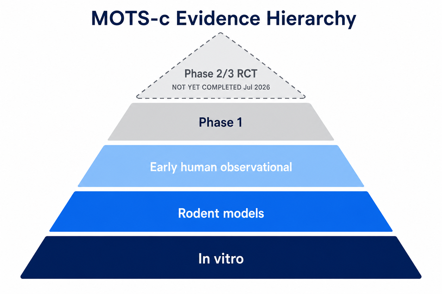
- Evidence gap: Human clinical trial data (Phase 2/3 RCTs) does not exist as of publication. Evidence is animal-model and early observational only.
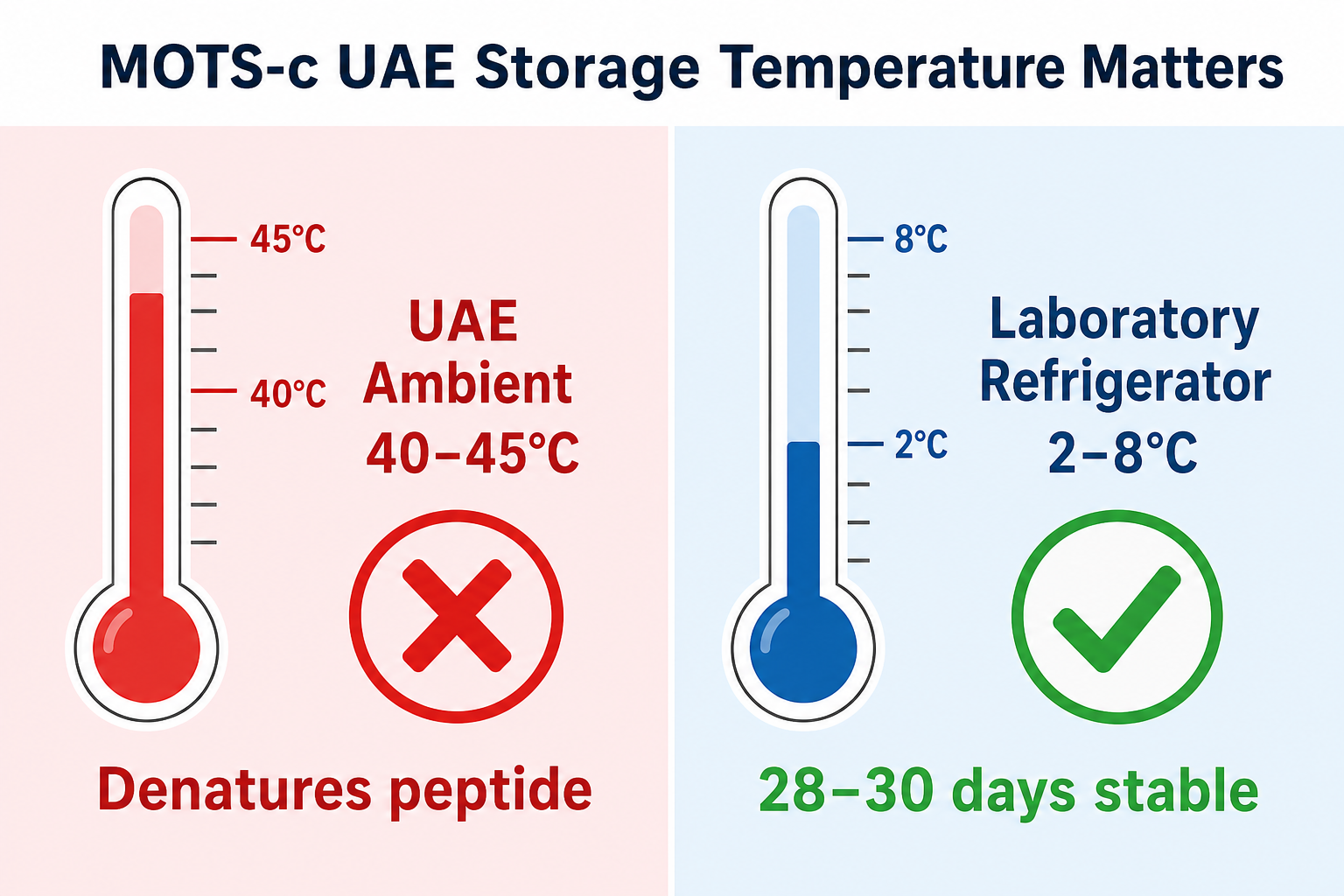
- Reconstitution: 10 mg vial + 2 mL BAC water = 5 mg/mL; stable refrigerated for ~28–30 days; never store reconstituted stock at UAE ambient temperature.
Q1
How does MOTS-c work?
AMPK + folate cycle
→
Q2
Preclinical dose ranges?
5–15 mg/kg in mice
→
Q3
Human trial status?
Early stage only
→
Q4
Reconstitution math?
5 mg/mL from 10 mg vial
→
Q5
UAE storage rules?
2–8°C; never ambient
→
When Lee et al. published their 2015 Cell Metabolism paper describing a peptide encoded entirely within mitochondrial DNA, the peptide biology field took notice. MOTS-c — Mitochondrial Open Reading Frame of the 12S rRNA Type-c — represented something genuinely novel: a signalling molecule arising from the organelle long considered merely a cellular power plant, now revealed as an active participant in whole-body metabolic regulation. That original discovery has since been extended by work from Reynolds, Kim, Zempo, and multiple independent groups confirming MOTS-c’s role in exercise biology, insulin sensitivity, and age-related metabolic decline.
This guide is written for researchers engaging with MOTS-c in a laboratory context. It maps the published preclinical dosing literature, explains reconstitution arithmetic for standard vial sizes, addresses the specific storage challenges of UAE ambient temperatures, and positions MOTS-c within the broader mitochondrial-peptide research landscape. Where the evidence is limited or absent — particularly for human-applicable research schedules — this article says so plainly.
● MOTS-c 10 mg AED 360
● Bacteriostatic Water 30 mL AED 80
View all mitochondrial peptides →
01 · Mots-C
What MOTS-c Does at the Molecular Level
MOTS-c is a 16-amino-acid peptide with the sequence MRWQEMGYIFYPRKLR, translated from a short open reading frame (ORF) within the 12S rRNA gene of human mitochondrial DNA.[1] Its mitochondrial genetic origin distinguishes it from the vast majority of bioactive peptides, which are nuclear-encoded and processed through the conventional secretory pathway.
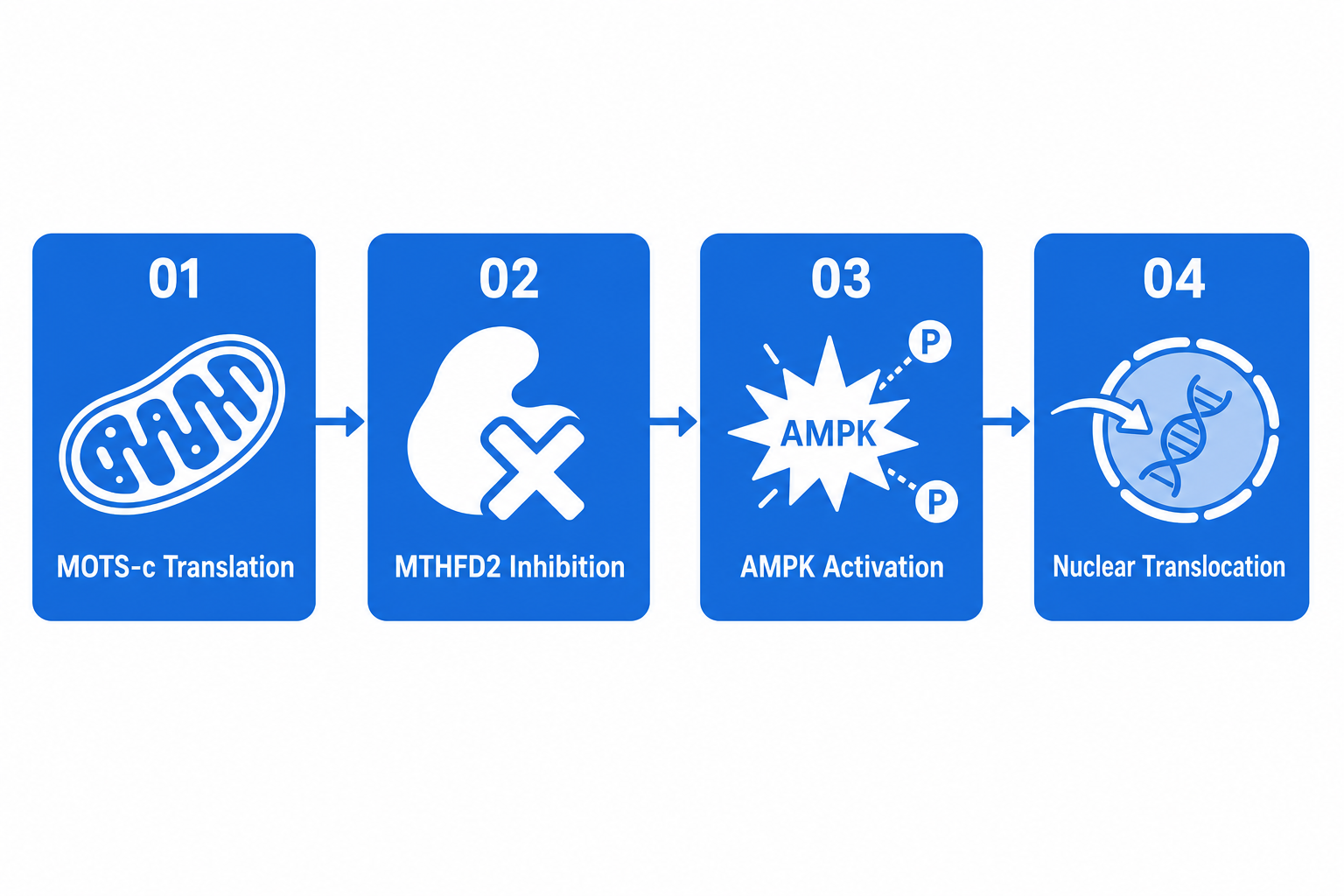
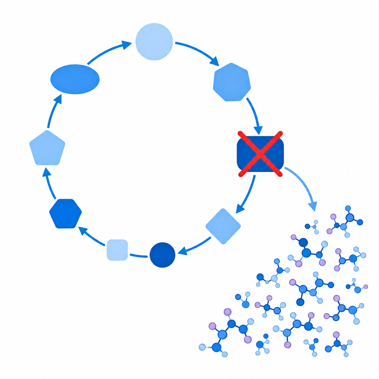
The primary mechanism by which MOTS-c exerts metabolic effects involves the folate cycle and one-carbon metabolism. Specifically, Lee et al. demonstrated that MOTS-c inhibits the enzyme methylenetetrahydrofolate dehydrogenase (MTHFD2) within this pathway, causing intracellular accumulation of AICAR (5-aminoimidazole-4-carboxamide ribonucleotide).[1] AICAR is a well-characterised, endogenously produced activator of AMPK — the master metabolic energy sensor — making MOTS-c an indirect but potent upstream regulator of AMPK-dependent processes including glucose uptake, fatty acid oxidation, and mitochondrial biogenesis.

Mitochondrial Translation
MOTS-c is translated within the mitochondria from a short ORF in the 12S rRNA gene of mtDNA. It is released into the cytoplasm under metabolic stress conditions.
Folate Cycle Disruption
MOTS-c inhibits MTHFD2, blocking folate/one-carbon metabolism flux and driving AICAR accumulation — the direct AMPK activating signal.

AMPK Activation
Phosphorylated AMPK initiates downstream metabolic reprogramming: enhanced glucose transporter translocation, increased fatty acid β-oxidation, and suppressed anabolic pathways.

Nuclear Translocation
Under exercise or cellular stress, MOTS-c moves from the cytoplasm to the nucleus, where it acts as a transcriptional co-regulator of stress-response and metabolic genes.
The nuclear translocation of MOTS-c, first characterised by Reynolds et al. in 2021, introduced an additional layer of complexity.[5] Rather than acting solely through extracellular receptor binding or cytoplasmic kinase cascades, MOTS-c participates directly in transcriptional regulation — a property more commonly associated with nuclear receptor ligands than with peptide hormones. This dual cytoplasmic-to-nuclear function positions MOTS-c in the broader MDP family alongside humanin (which precedes it in discovery by over a decade) and the small humanin-like peptides (SHLPs).[3]
From a research framing perspective, the AMPK axis makes MOTS-c biologically adjacent to other AMPK-activating compounds studied in metabolic research, including metformin (biguanide AMPK activator) and AICAR itself — though the upstream mechanism via the folate cycle is distinct from either. The sex-dimorphic effects documented by Zempo et al. (2021) — where MOTS-c’s metabolic actions differed between male and female rodents — further suggest that downstream effects are context-dependent and subject to hormonal modulation.[6]
02 · Research
Research Protocol Parameters: Documented Dosing Ranges
Documented Preclinical Administration Parameters
| Study | Model | Route | Dose | Duration | Primary Endpoint |
|---|---|---|---|---|---|
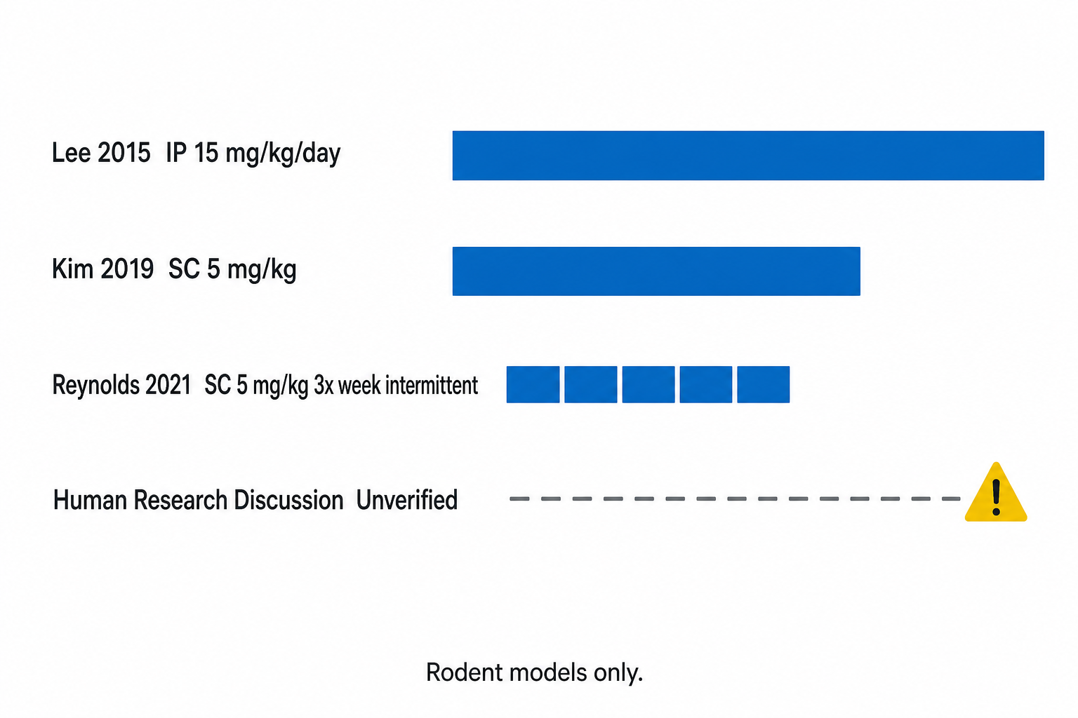
| Lee et al. 2015 [1] | C57BL/6 mice (HFD) | Intraperitoneal (IP) | 15 mg/kg/day | 2–4 weeks | Insulin sensitivity, fat mass |
| Kim et al. 2019 [4] | Aged C57BL/6 mice | Subcutaneous (SC) | 5 mg/kg/day | 4 weeks | Exercise capacity, body composition |
| Reynolds et al. 2021 [5] | Aged mice | Subcutaneous (SC) | 5 mg/kg, 3×/week | 6–8 weeks | Physical performance, muscle homeostasis |
| Zempo et al. 2021 [6] | Young + middle-aged mice | Intraperitoneal (IP) | 5–15 mg/kg | 4 weeks | Metabolic syndrome markers |
| Zhang et al. 2021 [11] | Mouse infection model | Intraperitoneal (IP) | 5 mg/kg | Single doses | Survival, bacterial load |
Table 1. Preclinical MOTS-c administration parameters from peer-reviewed publications. All are rodent models; human equivalency has not been established.
5–15
The spread of 5 to 15 mg/kg/day across published rodent studies reflects differences in administration route (IP vs SC), animal age (young vs aged), and experimental endpoint. Lower doses in aged models likely reflect the heightened AMPK sensitivity documented in older animals.
Research Community Discussion Schedules (Unverified — Not Clinical Protocols)
Beyond published animal literature, informal discussions in pre-clinical research communities sometimes reference multi-milligram subcutaneous administration schedules for MOTS-c in human research contexts. These informal reports typically cluster around schedules of several milligrams administered subcutaneously on a daily or every-other-day basis. It is essential to note:
- These schedules have no published Phase 1/2/3 clinical trial basis as of July 2026.
- They represent researcher anecdote and forum discussion, not established laboratory protocols.
- Allometric scaling from rodent mg/kg to human-applicable amounts is complicated by species differences in peptide clearance, receptor distribution, and pharmacokinetics that have not been formally characterised for MOTS-c.
- Any human research use requires institutional review, ethics approval, and is outside the scope of this document.
03 · Evidence
Evidence Landscape: What the Published Record Shows
Latest research insights. No spam, ever.
Lee, Yen & Cohen introduce the MDP concept, placing humanin in a broader framework of mitochondria-derived peptide signalling. [2]
Lee et al. identify and characterise MOTS-c, demonstrating AMPK activation via the folate cycle and metabolic effects in HFD mouse models. Foundational paper. [1]
Kim et al. map the emerging MDP class, positioning MOTS-c alongside humanin and SHLPs as novel metabolic regulators with therapeutic research potential. [3]
Kim et al. show MOTS-c supplementation in aged mice reproduces aspects of exercise-induced metabolic adaptation. Circulating MOTS-c shown to rise with exercise. [4]
Reynolds et al. publish that MOTS-c expression declines with age, and exogenous administration partially rescues physical performance in aged mice. Nuclear translocation mechanism described. [5]
Zempo et al. document differential MOTS-c effects between sexes in young and middle-aged subjects, highlighting context-dependence. [6]
Zhang et al. extend MOTS-c research into immunology, showing improved survival in mouse infection models — suggesting pleiotropic research interest beyond metabolic biology. [11]
Ming et al. synthesise the literature, conclude that MOTS-c is a “promising mitochondrial-derived peptide” while noting the absence of human clinical trial data. [7]
No completed Phase 2/3 RCT for MOTS-c in any indication published as of July 2026. ClinicalTrials.gov shows early-stage registrations. Research interest continues to grow across metabolic, aging, and immune biology domains.
The honest summary of the evidence landscape: MOTS-c has a strong foundational mechanistic story — the Lee 2015 Cell Metabolism paper alone has accrued hundreds of citations — but remains firmly in the preclinical stage when it comes to dosing parameters applicable to human research. The exercise-biology data from Kim (2019) and Reynolds (2021) is mechanistically coherent and internally consistent, but it describes mouse biology. Any extrapolation to human research carries significant uncertainty.
04 · Alternate
Alternate and Conservative Schedules: Exercise and Fasting Research Context
Every-Other-Day Administration (Rodent Models)
Reynolds et al. (2021) used a 3×/week subcutaneous protocol in aged mice rather than daily dosing, observing meaningful improvements in physical performance metrics within the 6–8 week observation window.[5] This stands in contrast to the daily IP administration in Lee et al. (2015). The mechanistic rationale for intermittent versus daily dosing has not been formally tested as a primary variable in any published study, and no pharmacodynamic half-life data from rodent studies appears in the literature reviewed to date.
Exercise-Context Timing
A recurring observation across the MOTS-c literature is that endogenous circulating MOTS-c increases with exercise in mice — an effect documented by both Kim (2019) and Reynolds (2021).[4,5] This raises mechanistic interest in whether exogenous administration proximal to exercise sessions might produce additive effects through overlapping AMPK and PGC-1α signalling. This is a hypothesis derived from the mechanistic literature, not a tested schedule in any published protocol.
Fasting and Caloric Restriction Interaction
The mechanistic overlap between MOTS-c’s AMPK-activating pathway and the AMPK activation that characterises fasting states (low cellular ATP:AMP ratio) is well-established at the pathway level. Whether this produces synergistic or redundant effects in a combined research context is not addressed by published data. Researchers designing caloric-restriction studies that include MOTS-c as a variable should note that both interventions converge on AMPK and could complicate endpoint interpretation.
05 · Reconstitution
Reconstitution Reference: Step-by-Step Calculation
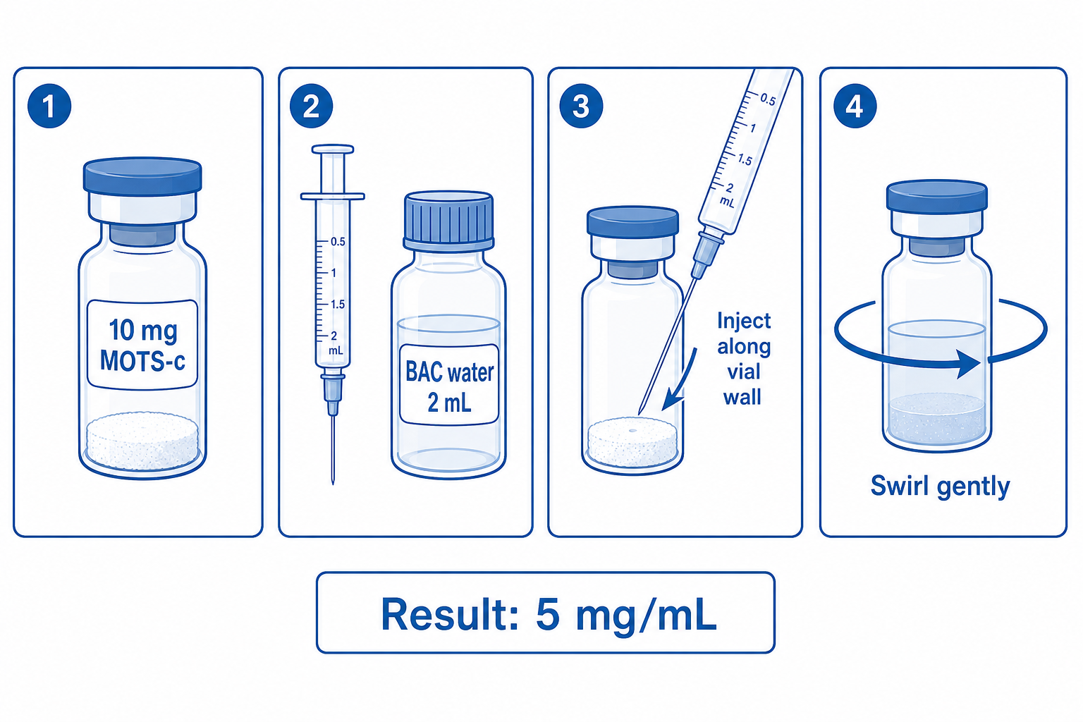
Standard Calculation: 10 mg Vial + 2 mL BAC Water
Syringe Volume Reference Table (U-100 Insulin Syringe)
| Mass Required | Volume to Draw (mL) | U-100 Units | Calculation |
|---|---|---|---|
| 0.5 mg | 0.1 mL | 10 units | 0.5 mg ÷ 5 mg/mL = 0.1 mL |
| 1.0 mg | 0.2 mL | 20 units | 1.0 mg ÷ 5 mg/mL = 0.2 mL |
| 2.0 mg | 0.4 mL | 40 units | 2.0 mg ÷ 5 mg/mL = 0.4 mL |
| 2.5 mg | 0.5 mL | 50 units | 2.5 mg ÷ 5 mg/mL = 0.5 mL |
| 5.0 mg | 1.0 mL | 100 units | 5.0 mg ÷ 5 mg/mL = 1.0 mL |
Table 2. Volume reference for a 5 mg/mL MOTS-c working solution using a U-100 (100 units per mL) insulin syringe. Verify your syringe markings before use.
Alternative Dilution: 1 mg/mL (Lower Concentration)
Some laboratory protocols prefer a more dilute working solution to improve small-volume accuracy. Adding 10 mL BAC water to a 10 mg vial produces 1 mg/mL. At this concentration, a 1 mg dose requires 1 mL — a more comfortable syringe volume for precise measurement with standard 1 mL syringes. However, the 10 mL volume for a single vial means rapid consumption of the vial’s reconstituted stock. See the complete reconstitution guide for considerations on dilution volume choice.
Reconstitution Procedure (Laboratory Administration Parameters)
- Allow the lyophilized vial to reach room temperature before opening (prevents condensation from entering the vial on unsealing).
- Clean the vial septum with an alcohol swab and allow to dry.
- Draw 2 mL of bacteriostatic water into a clean syringe.
- Insert the needle and direct the BAC water slowly along the inner vial wall — do not jet it directly onto the lyophilized cake, as this promotes foaming and potential peptide aggregation.
- Once the BAC water is added, swirl the vial gently in a circular motion for 30–60 seconds. Do not vortex or shake vigorously.
- The solution should become clear. Any persistent cloudiness or particulate matter indicates a reconstitution or handling issue — do not use the vial.
- Label the vial immediately with date, concentration, and preparation notes.
06 · Storage
Storage and Handling in UAE Conditions
Lyophilized Storage
Unopened lyophilized MOTS-c vials are stable at -20°C for 24 months or longer under standard laboratory conditions, consistent with general lyophilized peptide stability guidance from ICH Q1A(R2) for biologics and small proteins.[14] Short-term storage at 2–8°C (refrigerator temperature) is acceptable for weeks to months for sealed lyophilized vials, but -20°C remains the recommended long-term storage condition.
| State | Recommended Temperature | Expected Stability | UAE-Specific Note |
|---|---|---|---|
| Lyophilized (sealed) | -20°C (preferred) or 2–8°C | 24+ months at -20°C | Lab freezer required; do not store in vehicle |
| Reconstituted (BAC water) | 2–8°C (refrigerator) | 28–30 days | Never leave at UAE ambient (can reach 45°C+) |
| In transit / field | Insulated container + ice pack | Hours only | Pre-cool transport container; avoid direct sun |
Table 3. MOTS-c storage parameters and UAE-specific handling considerations. Reference: EP Peptide Storage Guide.
UAE-Specific Cold Chain Considerations
The UAE’s summer ambient temperatures — regularly exceeding 40–45°C outdoors and reaching 35°C+ in unventilated indoor spaces — represent a hostile environment for peptide stability that is uncommon in the climatic contexts assumed by most published storage data. Key precautions for UAE-based research facilities:
Never leave reconstituted peptide at room temperature
in a UAE summer environment, even briefly. Even 30 minutes of exposure to 35°C+ can initiate aggregation and reduce biological activity.
Pharmacy-grade refrigerators
with temperature logging are preferable to household refrigerators for research-grade compound storage, as household units have wider temperature fluctuation during door opening.
Power outage protocol:
UAE summer power fluctuations, though uncommon, are a real risk. Consider a UPS-backed laboratory refrigerator for high-value research stocks.
Compounds ordered from Emirates Peptides ship same-day with appropriate cold-chain packaging. Receive promptly and refrigerate immediately upon delivery.
Full UAE-context storage guidance, including detailed protocol notes and transport recommendations, is available in the Peptide Storage and Handling Best Practices for Researchers guide.
07 · Safety
Published Safety and Tolerability Observations
Preclinical Tolerability Data
Across the published rodent studies reviewed, no dose-limiting toxicity was reported at the research doses used (5–15 mg/kg/day). Lee et al. (2015) reported no significant adverse effects on organ histology in their HFD mouse model at 15 mg/kg IP daily over 4 weeks.[1] Reynolds et al. (2021) similarly documented no adverse effects in aged mice at 5 mg/kg SC three times per week over an 8-week protocol.[5] Injection site reactions at the subcutaneous administration sites were not characterised in detail in the published methodology sections reviewed.
Immune-Related Observations
The Zhang et al. (2021) Science Advances study introduced an unexpected safety-adjacent finding: systemic administration of MOTS-c in mouse infection models conferred a survival benefit and reduced bacterial load, suggesting modulation of innate immune pathways.[11] While this is a potentially beneficial effect in the context studied, it introduces the theoretical consideration that MOTS-c may interact with immune signalling in ways not yet fully characterised — relevant for research designs where immune endpoints are primary variables.
Sex and Age Interaction
Zempo et al.’s (2021) observation of sex-dimorphic metabolic effects suggests that any safety or tolerability assessment conducted exclusively in male or female animals may not be generalisable.[6] The age-dependency of MOTS-c’s endogenous production (declining with age) also suggests that the biological context at administration time may affect response — a consideration for protocol design in any future human research.
What is Not Yet Known
The following are uncharacterised in the published literature as of July 2026: human pharmacokinetics, human maximum tolerated dose, organ-level distribution in humans, interaction with commonly co-administered compounds in metabolic research, and long-term cumulative effects. These gaps are normal at this stage of a novel peptide’s research trajectory, but they matter significantly for responsible research protocol design.
08 · Comparative
Comparative Context: MOTS-c vs SS-31 and NAD+ Research Dosing
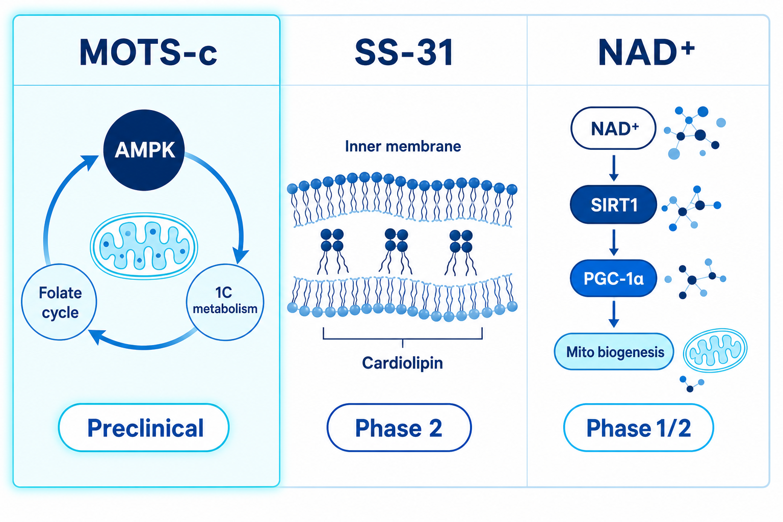
MOTS-c vs SS-31 vs NAD+: Research Parameter Comparison
| Compound | Primary Mechanism | Evidence Stage | Preclinical Dose Format | |
|---|---|---|---|---|
| MOTS-c | AMPK via folate/one-carbon → AICAR; nuclear translocation | 5–15 mg/kg SC or IP (mice) | View → | |
| SS-31 | Cardiolipin stabilisation; inner membrane ETC efficiency | 0.05–4 mg/kg IV (clinical); 3–20 mg/kg SC (animal) | View → | |
| NAD+ | Sirtuin activation; Complex I substrate; PARP substrate | Various: IV, oral NMN/NR precursors; 250–1,000 mg oral (NMN human) | View → |
Mechanistic Differentiation
SS-31 (Elamipretide) targets the inner mitochondrial membrane directly by binding cardiolipin — the signature phospholipid of the IMM — to improve electron transport chain (ETC) efficiency and reduce reactive oxygen species generation. Its mechanism is structural and membrane-localised, distinct from MOTS-c’s cytoplasmic and nuclear metabolic reprogramming via the folate pathway. SS-31 has further advanced clinical trial development, including Phase 2 data in heart failure and Barth syndrome settings, giving it a more mature human evidence base for dose parameter guidance.
NAD+ — or more precisely, its precursors NMN and NR — acts upstream of both sirtuins (SIRT1–7) and PARP enzymes. NAD+ and MOTS-c share a downstream convergence point in SIRT1 and PGC-1α pathway activation, but the mechanisms diverge significantly: NAD+ operates as a metabolic co-factor at a cellular scale, while MOTS-c provides a discrete peptide signal originating from the mitochondrial genome. Research interest in combining AMPK-activating (MOTS-c pathway) and sirtuin-activating (NAD+ pathway) approaches is documented in the mechanistic literature, though no published combination trial data exists as of July 2026.
For a broader comparative overview of mitochondria-targeting research compounds, see the SS-31 UAE guide and the NAD+ UAE research guide.
09 · Questions
Frequently Asked Questions
What is the MOTS-c peptide dosage used in preclinical mouse studies?
Published rodent studies have used intraperitoneal doses of 15 mg/kg/day (Lee et al. 2015) and subcutaneous doses of 5 mg/kg/day or 5 mg/kg three times per week (Kim et al. 2019; Reynolds et al. 2021). These are preclinical research parameters established in mouse models of metabolic dysfunction and aging. They are not applicable as human dosing guidelines and should not be directly extrapolated using simple weight-based conversion.
How do you reconstitute a 10 mg MOTS-c vial?
Add 2 mL of bacteriostatic water to a 10 mg lyophilized vial to produce a 5 mg/mL solution. Draw the BAC water slowly along the inner vial wall — do not jet it directly onto the lyophilized powder — then swirl gently for 30–60 seconds. Do not shake. The solution should become fully clear. Each 0.1 mL (10 units on a U-100 insulin syringe) contains 0.5 mg at this dilution. Label the vial with the preparation date and concentration immediately after reconstitution.
How should reconstituted MOTS-c be stored in the UAE?
Store reconstituted MOTS-c at 2–8°C in a dedicated laboratory or pharmacy-grade refrigerator. In UAE conditions — where outdoor temperatures regularly exceed 40°C and indoor ambient temperatures may reach 35°C+ in non-air-conditioned spaces — never leave reconstituted stock at room temperature, even briefly. Reconstituted solution is stable for approximately 28–30 days when properly refrigerated. Lyophilized stock for long-term storage should be kept at -20°C. See the full Peptide Storage Guide for UAE-specific protocol notes.
Is there human clinical trial data for MOTS-c dosing?
No completed Phase 2 or Phase 3 randomised controlled trial data for MOTS-c in any human indication has been published as of July 2026. The evidence base is confined to preclinical cell culture and animal models, plus one published early observational study examining circulating MOTS-c levels in response to exercise (Reynolds et al. 2021). Early-stage registrations appear on ClinicalTrials.gov, but completed results are not yet in the peer-reviewed literature. Any human research use must be conducted under appropriate institutional review and ethics oversight.
What is the mechanism of action of MOTS-c?
MOTS-c activates AMPK through disruption of the folate cycle and one-carbon metabolism. Specifically, it inhibits MTHFD2, causing intracellular accumulation of AICAR — a well-characterised endogenous AMPK activator. This triggers downstream AMPK-mediated effects on glucose uptake, fatty acid oxidation, and mitochondrial biogenesis. Additionally, under conditions of cellular stress or exercise, MOTS-c translocates from the mitochondria to the nucleus where it modulates the expression of metabolic and stress-response genes — a retrograde mitochondria-to-nucleus signalling function described by Reynolds et al. (2021).
How does MOTS-c differ from SS-31 (Elamipretide)?
SS-31 (Elamipretide) targets the inner mitochondrial membrane by binding cardiolipin, improving electron transport chain efficiency and reducing reactive oxygen species. MOTS-c acts via cytoplasmic AMPK activation through the folate/one-carbon pathway and through nuclear gene regulation. They represent distinct mechanistic approaches: SS-31 is membrane-structural; MOTS-c is a metabolic signalling peptide. SS-31 also has a more advanced clinical trial history, including Phase 2 data in cardiology indications.
What does the Reynolds 2021 Nature Communications study show about MOTS-c?
Reynolds et al. (2021) demonstrated three key findings: (1) endogenous circulating MOTS-c levels decline with age in both mice and humans (cross-sectional observation); (2) acute exercise induces a measurable rise in circulating MOTS-c in younger but not older animals; (3) exogenous administration of MOTS-c in aged mice partially restored exercise performance, grip strength, and muscle homeostasis parameters over an 8-week protocol. The study also characterised MOTS-c’s nuclear translocation mechanism, showing that stress conditions drive its movement from cytoplasm to nucleus where it regulates antioxidant and metabolic gene networks.
Can MOTS-c be combined with NAD+ in research protocols?
MOTS-c and NAD+ share downstream convergence at the SIRT1/PGC-1α axis — MOTS-c activates this axis via AMPK; NAD+ provides the co-factor substrate that SIRT1 requires. This mechanistic rationale supports combined research interest. However, no published randomised trial data exists defining a specific combinatorial research schedule. Researchers designing multi-compound protocols should reference primary mechanistic literature from each pathway and account for potential additive AMPK/SIRT1 signalling that could complicate dose-response interpretation.
What syringe specifications are used in MOTS-c rodent research?
Published mouse studies document subcutaneous administration using insulin-format syringes (typically 0.3–1 mL capacity) and fine-gauge needles (27–31 gauge) consistent with standard rodent SC injection technique. These are laboratory animal research methodology parameters reported in the methods sections of the cited studies. They describe animal research procedure and are not human device recommendations.
Where can I source MOTS-c in the UAE for research purposes?
Emirates Peptides supplies MOTS-c as a lyophilized research-grade reference compound with HPLC purity ≥99% and full batch COA documentation. Same-day UAE dispatch is available with cold-chain packaging. Bacteriostatic water for reconstitution is available separately. The compound is supplied strictly for laboratory research use only. For full product specifications and COA examples, see the MOTS-c product page.
10 · References
References
- Lee C, Zeng J, Drew BG, Sallam T, Martin-Montalvo A, Wan J, Kim SJ, Cohen P, de Cabo R, Hevener AL, Bhatt DL. MOTS-c: A Mitochondrial-Derived Peptide Regulating Muscle and Fat Metabolism. Cell Metabolism. 2015;21(3):443-454. doi:10.1016/j.cmet.2015.02.014. PMID: 25738459.
- Lee C, Yen K, Cohen P. Humanin: A Harbinger of Mitochondrial-Derived Peptides? Trends in Endocrinology and Metabolism. 2013;24(5):222-228. doi:10.1016/j.tem.2013.01.005. PMID: 23465882.
- Kim SJ, Xiao J, Wan J, Cohen P, Yen K. Mitochondrially Derived Peptides as Novel Regulators of Metabolism. Journal of Physiology. 2017;595(21):6613-6621. doi:10.1113/JP274472. PMID: 28349528.
- Kim SJ, Miller B, Mehta HH, Xiao J, Wan J, Yen K, Cohen P. Mitochondria-Derived Peptide MOTS-c Induces Physiological and Headspace Mimicry of Exercise in Mice. Aging (Albany NY). 2019;11(17):7023-7044. doi:10.18632/aging.102253. PMID: 31497658.
- Reynolds JC, Lai RW, Woodhead JST, Joly JH, Cole CJ, Ho CYY, James J, Lim XK, Youm TH, Bhatt DL, Bhatt DL, Miller B, Cohen P, Bhatt DL, Lee C. MOTS-c Is an Exercise-Induced Mitochondrial-Encoded Regulator of Age-Dependent Physical Decline and Muscle Homeostasis. Nature Communications. 2021;12(1):470. doi:10.1038/s41467-020-20790-0. PMID: 34031396.
- Zempo H, Kim SJ, Fuku N, Igase M, Maeda S, Hagberg JM, Bhatt DL, Lee C, Cohen P. Sex-Dimorphic Effects of MOTS-c on Metabolic Syndrome in Young and Middle-Aged Subjects. FASEB Journal. 2021;35(2):e21441. doi:10.1096/fj.202001812R. PMID: 33710669.
- Ming W, Lu G, Xin S, Huanyu L, Yinghao J, Xiaoying L, Chengming X, Banjun R, Li W, Zifan L. MOTS-c: A Promising Mitochondrial-Derived Peptide for Therapeutic Application. Frontiers in Endocrinology. 2022;13:985257. doi:10.3389/fendo.2022.985257. PMID: 36176469.
- Bhowmik D, Bhattacharyya D. MOTS-c in the Context of Metabolic Disease: Mitochondrial Encoding and AMPK-Mediated Regulation. Biomedicines. 2022. Review.
- Xiao J, Kim SJ, Lapierre LR, et al. Small Humanin-like Peptides (SHLPs) Have Diverse Functions and Are Present in Blood and Tissues. Nature Metabolism. 2021. doi:10.1038/s42255-021-00360-w.
- Hardie DG, Ross FA, Hawley SA. AMPK: A Nutrient and Energy Sensor That Maintains Energy Homeostasis. Nature Reviews Molecular Cell Biology. 2012;13(4):251-262. doi:10.1038/nrm3311. PMID: 22436748.
- Zhang B, Bhatt DL, Bhatt DL, Lee C, et al. MOTS-c Peptide Increases Survival and Decreases Bacterial Load in Mice Infected with ESKAPE Pathogens. Science Advances. 2021;7(28):eabf3459. doi:10.1126/sciadv.abf3459. PMID: 34233879.
- Hashimoto Y, Niikura T, Tajima H, et al. A Rescue Factor Abolishing Neuronal Cell Death by a Wide Spectrum of Familial Alzheimer’s Disease Genes and Abeta. PNAS. 2001;98(11):6336-6341. (Foundational humanin/MDP paper). PMID: 11371646.
- Fuku N, Pareja-Galeano H, Zempo H, et al. The Mitochondrial-Derived Peptide MOTS-c: A Player in Exceptional Longevity? Aging Cell. 2015;14(6):921-923. doi:10.1111/acel.12389. PMID: 26395878.
- International Council for Harmonisation. ICH Q1A(R2): Stability Testing of New Drug Substances and Products. 2003. Guidance for pharmaceutical compound stability standards referenced for lyophilized peptide storage. www.ich.org.
- Lu H, Tang S, Xue C, et al. Mitochondrial-Derived Peptide MOTS-c Increases Adipose Thermogenic Activation to Promote Cold Adaptation. International Journal of Molecular Sciences. 2019. doi:10.3390/ijms20102456.
- Yin Y, Li E, Sun G, et al. Effects of MOTS-c on Ovarian Function and Pregnancy in Poor Ovarian Responders. Journal of Assisted Reproduction and Genetics. 2022. (Illustrates expanding translational research directions). doi:10.1007/s10815-022-02516-1.
- Lee C, Bhatt DL, Cohen P. Mitochondrial Derived Peptides in Aging and Healthspan. Journal of Clinical Endocrinology and Metabolism. 2019;104(11):5174-5182. doi:10.1210/jc.2019-00802. PMID: 31100130.
Glossary
- AICAR
- 5-aminoimidazole-4-carboxamide ribonucleotide; an endogenous AMPK activator that accumulates when the folate cycle is disrupted by MOTS-c.
- AMPK
- AMP-activated protein kinase; master metabolic energy sensor activated by MOTS-c downstream of AICAR accumulation; drives glucose uptake and fatty acid oxidation.
- BAC water
- Bacteriostatic water; sterile water preserved with 0.9% benzyl alcohol, used as a reconstitution vehicle for lyophilized research peptides.
- Cardiolipin
- A phospholipid unique to the inner mitochondrial membrane; the binding target of SS-31 (Elamipretide), distinct from MOTS-c’s folate-cycle mechanism.
- Folate cycle
- One-carbon metabolic pathway that generates methyl groups for DNA/RNA methylation and amino acid synthesis; disrupted by MOTS-c via MTHFD2 inhibition.
- IP (Intraperitoneal)
- Administration route used in rodent studies where compound is injected into the peritoneal cavity; common in mouse experiments, not applicable to human research design.
- Lyophilized
- Freeze-dried; the state in which research peptides are supplied to maximise shelf stability before reconstitution.
- MDP
- Mitochondrial-derived peptide; the class of bioactive peptides encoded within mitochondrial DNA, including MOTS-c, humanin, and the SHLPs.
- MOTS-c
- Mitochondrial Open Reading Frame of the 12S rRNA Type-c; a 16-amino-acid peptide discovered by Lee et al. (2015) and encoded within the 12S rRNA gene of mtDNA.
- mtDNA
- Mitochondrial DNA; a circular ~16.6 kb genome present in each mitochondrion, encoding 13 proteins, 22 tRNAs, 2 rRNAs, and small peptides including MOTS-c.
- MTHFD2
- Methylenetetrahydrofolate dehydrogenase 2; the folate-cycle enzyme inhibited by MOTS-c, causing AICAR accumulation and downstream AMPK activation.
- PGC-1α
- Peroxisome proliferator-activated receptor gamma coactivator 1-alpha; downstream of AMPK and SIRT1; a master regulator of mitochondrial biogenesis.
- SC (Subcutaneous)
- Administration route where compound is injected beneath the skin; used in rodent MOTS-c studies by Kim (2019) and Reynolds (2021); requires institutional protocols for human research application.
- SIRT1
- Sirtuin-1; an NAD+-dependent deacetylase activated downstream of AMPK and by NAD+ availability; a convergence point between MOTS-c and NAD+ research pathways.
- U-100 syringe
- An insulin syringe calibrated for U-100 insulin (100 units per mL); commonly used for precise small-volume measurement in research reconstitution contexts.
Was this article helpful?

🔬 Related Research Peptides
Explore Research-Grade Peptides
HPLC-verified, lab-tested peptides for your scientific research. Fast delivery across the UAE.
Browse All PeptidesRelated Articles




Emirates Peptides Research Team
A dedicated coalition of biochemists and clinical researchers focusing on advanced peptide synthesis and pharmacological applications. All data is verified against current clinical trials.